RESEARCH ARTICLE
Doi: 10.5578/tt.53947
Tuberk Toraks 2017;65(3):258-259

Three-dimensional ct scan for Swyer-James syndrome
Satoshi HAGIMOTO1, Gen OHARA1, Kunihiko MIYAZAKI1, Hiroaki SATOH1
1 Division of Respiratory Medicine, Mito Medical Center, Tsukuba University, Mito, Japan
1 Tsukuba ?niversitesi Mito Tıp Merkezi, Solunum B?l?m?, Mito, Japonya
To the Editor,
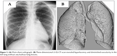
A 16-year-old woman was referred due to a left hyperlucent lung, which was incidentally observed on a chest radiograph (Figure 1A).The patient had a history of respiratory infection in her infancy. A three-dimensional (3-D) CT scan revealed hyperlucency and diminished vascularity in the left lung without any bullous lunglesions (Figure 1B). On the basis of these findings, the patient was diagnosed to have Swyer-James syndrome (1,2).When unilateral hyperlucent lung is discovered, a 3-D CT scan would provideimportant clinical information as observed in this case. Although extremely rare, Swyer-James syndrome should be included in the differential diagnosis of unilateral hyperlucent lungif patients have a history of pulmonary infection in their early childhood.
3-D CT scan would provide critical information in distinguishing between "Swyer-James syndrome" and "other diseases exhibiting unilateral hyperlucent lung". In addition, information obtained by 3-D CT scan would advance differential diagnosis without any other invasive examination, therefore, there would be benefits of low invasiveness and economic merit for patients.
REFERENCES
- Swyer PR, James GCW. A case of unilateral pulmonary emphysema.Thorax 1953;8:133-6.
- Marti-Bonmati L, Ruiz Perales F, Catala F, Mata JM, Calonge ECT findings in Swyer-James syndrome. Radiology 1989; 172:477-80.
Yazışma Adresi (Address for Correspondence)
Dr. Hiroaki SATOH
Division of Respiratory Medicine,
Mito Medical Center, Tsukuba University,
Mito - Japan
e-mail: hirosato@md.tsukuba.ac.jp
Geliş Tarihi/Received: 25.03.2017 - Kabul Ediliş Tarihi/Accepted: 31.03.2017
